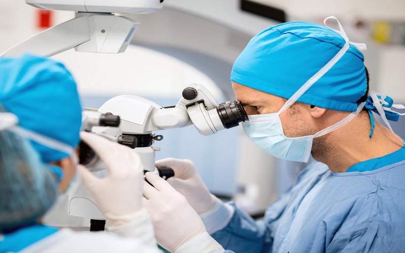
Welcome to the world of retina surgery. Here, delicate tasks carried out by skilled ophthalmologists help preserve and restore vision. It all starts with the Eye exam Brooklyn. This is the first step in diagnosing retinal problems. In this blog, we unravel the intricate procedures involved in retina surgery.
Understanding the Eye
To grasp retina surgery, you need to understand the eye. The retina is a thin layer at the back of the eye. It captures light and sends it to the brain. When it’s damaged, vision suffers.
The Need for Surgery
Surgery is the answer for some retinal problems. These include retinal detachment, tears or holes, and macular degeneration. It can help save or restore vision.
The Process of Surgery
Retina surgery is a detailed process. It usually involves one of three procedures: vitrectomy, scleral buckle, or pneumatic retinopexy. Vitrectomy removes the vitreous gel to access the retina. Scleral buckle supports the retina. Pneumatic retinopexy uses a gas bubble to repair the retina.
Preparation and Recovery
Before surgery, an eye exam is crucial. It allows the ophthalmologist to assess the retina’s condition. After surgery, recovery varies. It can involve some discomfort and adjustments. But overall, many patients see improvement in their vision.
| Procedure | Description |
| Vitrectomy | Removal of the vitreous gel to repair the retina |
| Scleral Buckle | A band is placed around the eye to support the retina |
| Pneumatic Retinopexy | A gas bubble is injected into the vitreous space to push the retina back into place |
Final Thoughts
Retina surgery is a complex process. It requires skill and precision. But it offers hope to those with retinal problems. With the right care, it can make a significant difference in vision and quality of life.
For more information on retina surgery, visit the National Center for Biotechnology Information.

